Home
/ Female Back Bones Diagram : Muscle Anatomy For Female And Male 2 Dry Erase Clipboard : Bones of the pelvis and lower back.
Female Back Bones Diagram : Muscle Anatomy For Female And Male 2 Dry Erase Clipboard : Bones of the pelvis and lower back.
Female Back Bones Diagram : Muscle Anatomy For Female And Male 2 Dry Erase Clipboard : Bones of the pelvis and lower back.. This shopping feature will continue to load items when the enter key is pressed. The red lines point individual bones and the names are writen in singular, the blue lines conect to group of bones and are in plural form. It represents a vestigial tail, hence the common term tailbone. For more anatomy content please follow us and visit our website: You can locate it behind your vaginal opening.
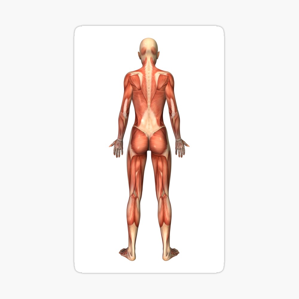
There are three parts to the trapezius. Diagram of a human female skeleton back view. Human internal organs vector vector isolated illustration of human internal organs in female body. The pubic bone is a joint where the two halves of the pelvis meet. The trapezius or trapezoid muscles are two paired muscles that extend from the base of the thoracic vertebrae in the spine to the occipital bone and run out to the spine of the scapula.
Anatomy Stock Illustrations 265 169 Anatomy Stock Illustrations Vectors Clipart Dreamstime from thumbs.dreamstime.com It represents a vestigial tail, hence the common term tailbone. This diagram depicts picture of the female body 744×992 with parts and labels. The vertebrae, which stack like spools of thread, support the back and protect the spinal cord. Muscle anatomy triceps 12 photos of the muscle anatomy triceps. Just need a glimpse, leave your valuable advice let us know , and subscribe us! This shopping feature will continue to load items when the enter key is pressed. The trapezius or trapezoid muscles are two paired muscles that extend from the base of the thoracic vertebrae in the spine to the occipital bone and run out to the spine of the scapula. The human spine is composed of 33 vertebrae that interlock with each other to form the spinal column.
At the back of each bone in the spine (vertebra) are bony points called processes, which muscles attach to.
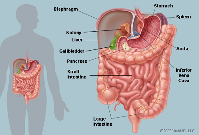
Related posts of human back bones diagram female pelvis bones images. The spine anatomy is a complex structure. Diagram chest muscles, diagram human back muscles, diagram of back muscles and bones, diagram of back muscles and ligaments, diagram of back muscles and nerves, diagram of back muscles pain, diagram of lower back muscles, diagram shoulder muscles, human muscles, diagram chest muscles, diagram. Diagram of a human female skeleton, back view. This diagram depicts picture of the female body 744×992 with parts and labels. 9 photos of the diagram of female back muscles. This article looks at the anatomy of the back, including bones, muscles, and nerves. The trapezius or trapezoid muscles are two paired muscles that extend from the base of the thoracic vertebrae in the spine to the occipital bone and run out to the spine of the scapula. Stomach, liver, intestine, bladder, lung, testicle, uterus, spine, pancreas, kidney, heart. Diagram of a human female skeleton back view. There are exactly 26 bones in the hand and 26 in the foot. Spinal vertebrae bone spine vertebra toracica spinal cord spine structure back diagram spine sections spinal cord vertebrae spinal structure health diagram. Bones make up about 14 percent of our body weight.
Human back bones diagram, find out more about human back bones diagram. This diagram depicts picture of the female body 744×992 with parts and labels. This shopping feature will continue to load items when the enter key is. Can you feel the bumps of your vertebrae along your back? The coccyx is a triangular arrangement of bone that makes up the very bottom portion of the spine below the sacrum.
Female Muscular System Back View Greeting Card By Stocktrekimages Redbubble from ih1.redbubble.net Women have a wider pelvis (hips) than men because the opening has to be wide enough to let a baby pass through when it's born. Sciatica medical health care vector illustration scheme with lower spine and sciatic nerve pain in leg. The pubic bone is a joint where the two halves of the pelvis meet. Our latest youtube film is ready to run. This diagram depicts skeletal images 744×1314 with parts and labels. The red lines point individual bones and the names are writen in singular, the blue lines conect to group of bones and are in plural form. The vertebral column of the lower back includes the five lumbar vertebrae, the sacrum, and the coccyx. The bones of the pelvis and lower back work together to support the body's weight, anchor the abdominal and hip muscles, and protect the delicate vital organs of the vertebral and abdominopelvic cavities.
At the back of each bone in the spine (vertebra) are bony points called processes, which muscles attach to.
The muscles of the lower back help stabilize, rotate, flex, and extend the spinal column, which is a bony tower of 24 vertebrae that gives the body structure and houses the spinal cord. Our latest youtube film is ready to run. The vertebral column of the lower back includes the five lumbar vertebrae, the sacrum, and the coccyx. Stomach, liver, intestine, bladder, lung, testicle, uterus, spine, pancreas, kidney, heart. This shopping feature will continue to load items when the enter key is. 4 photos of the diagram of a female lower back. The red lines point individual bones and the names are writen in singular, the blue lines conect to group of bones and are in plural form. Diagram of lower back and hips, diagram of lower back muscles and ligaments, diagram of lower back organs, lower back anatomy diagram, lower back diagram bones, lower back diagram pelvis, human anatomy, diagram of lower back and hips, diagram of lower back muscles and ligaments, diagram of lower. Back anatomy stock photos and images. This article looks at the anatomy of the back, including bones, muscles, and nerves. It also covers some common conditions and injuries that can affect the back. Human back bones diagram, find out more about human back bones diagram. This shopping feature will continue to load items when the enter key is pressed.
The spine anatomy is a complex structure. It represents a vestigial tail, hence the common term tailbone. You might also like this photos or back to diagram of female body. An illustration showing a theory of vision published in treatise on man by rene descartes. The thigh bone (femur) is the longest bone in the body.
The Abdomen Human Anatomy Picture Function Parts Definition And More from img.webmd.com The spine diagram shown below, consists of many bones or vertebrae,soft discs,the spinal cord, and spinal nerves. See lumbar spine anatomy diagram stock video clips. A tough, springy disc of cartilage sits between the vertebrae of your spine. Diagram of lower back and hips, diagram of lower back muscles and ligaments, diagram of lower back organs, lower back anatomy diagram, lower back diagram bones, lower back diagram pelvis, human anatomy, diagram of lower back and hips, diagram of lower back muscles and ligaments, diagram of lower. The pubic bone is a joint where the two halves of the pelvis meet. The muscles of the lower back help stabilize, rotate, flex, and extend the spinal column, which is a bony tower of 24 vertebrae that gives the body structure and houses the spinal cord. For more anatomy content please follow us and visit our website: The bones of the pelvis and lower back work together to support the body's weight, anchor the abdominal and hip muscles, and protect the delicate vital organs of the vertebral and abdominopelvic cavities.
An illustration showing a theory of vision published in treatise on man by rene descartes.
Diagram chest muscles, diagram human back muscles, diagram of back muscles and bones, diagram of back muscles and ligaments, diagram of back muscles and nerves, diagram of back muscles pain, diagram of lower back muscles, diagram shoulder muscles, human muscles, diagram chest muscles, diagram. Female muscle groups anatomical fitness vector illustration, sports training informative chart. Flexibility especially in the lower back and neck allowing us to bend and twist in a full variety of movements strength provided by the bones discs joints and supportive muscles and connective tissue that allows us to stand upright and move about with precision. Touch device users can explore by touch or with swipe gestures. Muscle anatomy triceps 12 photos of the muscle anatomy triceps. The bones of the superior portion of the skull are known as the cranium and protect the brain from damage. You might also like this photos or back to diagram of female body. Just need a glimpse, leave your valuable advice let us know , and subscribe us! This diagram depicts picture of the female body 744×992 with parts and labels. It also covers some common conditions and injuries that can affect the back. The trapezius or trapezoid muscles are two paired muscles that extend from the base of the thoracic vertebrae in the spine to the occipital bone and run out to the spine of the scapula. The spinal cord itself is about 45 cm (18 in) in men and 43 cm (17 in) long in women. There are three parts to the trapezius.
